Posts Tagged ‘Breast Cancer’
Is there a cure for cancer? You bet there is! Part 5

Dr. Virginia Livingston who found a cure for cancer
As we near the end of the current series on the cancer scam I want to introduce you to other experts that have either worked with Dr. Virginia Livingston or who believes in her invention/protocol.
One of the first that comes to mind is her dear associate and friend Dr. Alan Cantwell who can be seen with Dr. Livingston in the above photograph.
His expertise is second to non and in this part it would be appropriate to print a full interview carried out with Dr. Cantwell back in 2007 who also happens to give reference to many other fine cancer researcher, most of whom were never identified or given the chance to prove they had all found a combined cure for cancer and many other bacterium based diseases:
11 Sep 2007
Dr. Alan Cantwell has investigated the phenomenon of cancer bacteria for over thirty years. A graduate of New York Medical College, Cantwell completed a residency program in dermatology at Long Beach Veteran’s Administration Hospital in Long Beach, CA and then practiced in the dermatology department of Kaiser-Permanente in Hollywood, California, from 1965 until his retirement in 1994. Dr. Cantwell is the author of more than thirty published papers on breast cancer, lymphoma, Kaposi’s sarcoma, Hodgkin’s Disease, lupus, scleroderma, AIDS, and other immunological diseases. These papers have appeared in many peer-reviewed journals, including Growth, International Journal of Dermatology, Journal of Dermatologic Surgery and Oncology, and the Archives of Dermatology. He has also written The Cancer Microbe and Four Women Against Cancer and several books on AIDS.

Dr. Alan Cantwell
- How did you become interested in looking for bacteria, first in diseases like scleroderma and later in cancer?
It all started when I was a second year resident in dermatology. I was in the medical library and I came across a paper in the Southern Medical Journal describing a group of people who had been given allergy injections and who subsequently developed deep skin infection with tuberculosis-like germs. It was thought the allergy injection bottles were contaminated with these bacteria.
At the time, I had a mentally disturbed patient who had been given multiple injections of medications into her buttocks. She later developed deep painful skin nodules in the same areas. No one knew what was causing these nodules that were diagnosed as “panniculitis,” an inflammation of the fat layers of the skin. I thought, “Let’s culture a skin biopsy from one of these deep nodules and see if I can find any TB-like germs.” I was amazed when Eugenia Craggs, the technician at the TB lab, reported that “acid-fast” bacteria were discovered in the skin tissue. I thought “Hey this is just like the article!”
We also had three other patients with “panniculitis” of the fatty portion of the skin, all of unknown cause. I took biopsy samples and TB-like bacteria were found in all four. These cases were later reported in the Archives of Dermatologyin 1966. At the time my dermatology professor was J. Walter Wilson, who was also a world famous mycologist, an expert in fungal diseases. He was somewhat skeptical about my findings of acid-fast bacteria in all these four patients and he suggested I use a scleroderma patient as a “control.” Scleroderma is a so-called “collagen disease” where the skin becomes hardened. The disease can affect the internal organs and is sometimes fatal. The cause is unknown, and bacteria were never thought to cause this disease. Dr. Wilson said I should check a scleroderma skin biopsy because that would serve as a negative “control” case. I was astonished when Eugenia Craggs called me from the TB lab and told me the skin tissue grindings of the scleroderma sample were positive for acid-fast bacteria, the kind of bacteria found in tuberculosis. She would try and grow the germ in a TB culture. After much searching I was also able to find a few acid-fast rod forms of bacteria in the scleroderma skin biopsy microscopic sections prepared by the pathologist.
The scleroderma bacterial took a long time to grow and could not be diagnosed as a TB germ or other definite “atypical” mycobacteria. The microbe was highly pleomorphic (various forms). There were round staphylococccal forms, as well as typical acid-fast rod forms. Eventually this isolate became fungal-like and “actinomycete- like.” Despite expert opinion, it was impossible to classify the microbe into a specific species. This case of scleroderma was reported in The Archives of Dermatology in 1966.
Some time later, Roy Averill, one of the dermatology residents, told me he heard a woman physician being interviewed on a San Diego radio talk show. She was explaining how she found TB-like bacteria in scleroderma in the late 1940s. That woman was Virginia Livingston M.D. She quickly became a dear friend and mentor in my scleroderma research. She told me that scientists at the Pasteur Institute in Belgium also reported finding acid-fast bacteria in scleroderma in 1953, thus confirming her own research.
I naturally thought all these reports in the medical journals would be recognized by other dermatologists and scientists, and that scleroderma would be recognized as an infectious disease caused by acid-fast bacteria. But after more than a half-century, I’m sad to say that scleroderma is still considered a disease “of unknown etiology” and the bacteria we found are simply ignored. After discovering acid-fast bacteria in scleroderma, Livingston found similar bacteria in cancer. This made her one of the most controversial physicians in America, as detailed in my book, “The Cancer Microbe.”
- How did you identify the bacteria in your samples?
I began my dermatology practice at Kaiser in Hollywood in 1965. Virginia Livingston introduced me to Dan Kelso, a Los Angeles microbiologist who thereafter cultured my skin biopsy samples from scleroderma, and later from lupus erythematosus and a variety of cancers. Depending on the case, sometimes he cultured Staphylococcus epidermidis, or corynebacteria, more rarely streptococci, and pleomorphic bacteria that appeared sporadically as acid-fast bacteria similar to Mycobacterium tuberculosis.
Naturally I attempted to find acid-fast rod forms in my specially-stained skin biopsy sections, because these forms are the typical forms signifying infection with Mycobacterium tuberculosis or other species of mycobacteria. “Acid-fast” refers to red-stained mycobacteria that can be observed after staining tissue samples with a special procedure and a special dye. At first, I didn’t see the L-form bacteria since they react differently to acid staining. Instead of rod-forms, they appeared as round forms which were only partially acid-fast, staining purple or magenta with the acid-fast stain. It took me many years to finally realize that these partially acid-fast and round forms were bona fide growth forms of mycobacteria. The typical bright red-stained acid-fast rod forms of mycobacteria are unique and easily recognized by pathologists, but unfortunately the non-acid-fast round forms are not recognized and accepted by pathologists. For a long time I passed over these granular and “dusty” tiny forms as meaningless, not realizing that they were, in actuality, what L-forms look like!
I knew basically nothing about the microscopic appearance of L-form bacteria (also known as cell wall deficient bacteria and “mycoplasma”) until I carefully read the published papers of microbiologist Lida Mattman. Then I realized all the guises that bacteria can undergo, including transformation into “large bodies.” At that point, I went back and looked at my first case of scleroderma and realized that one skin biopsy sample contained large L-form bodies that appeared as yeast and fungal-like forms! These forms, in 1966, were dismissed as “fat degeneration” by one pathologist; and the biologist thought they looked like yeast cells.
These large L-forms are compatible with what pathologists recognize as Russell Bodies. William Russell (1852-1940) was a well-known Scottish pathologist who first discovered “the parasite of cancer” in 1890. His view of an infectious agent in cancer was dismissed in the early part of the twentieth century. However, I believe Russell bodies are actually large growth forms of cell wall deficient bacteria — and that Russell was indeed recognizing an infectious agent in cancer. More than a half-century later, Lida Mattman was able to transform mycobacteria into “large bodies” by exposing them to antibiotics. For more information on Russell and pictures of Russell bodies, Google my paper “The Russell Body” in the Journal of Independent Medical Research (joimr.org).
The fact that L-form bacteria have a “life cycle” and can appear in so many different shapes and sizes (pleomorphism) may be why they are so hard to eradicate and why the immune system cannot cope with them. Maybe the large Russell bodies are harder to kill. Or maybe they are easier to kill. I don’t know.
- You found bacteria in the tissues of people who died of certain cancers and AIDS and scleroderma at autopsy. What gave you the idea to look for bacteria in autopsies?
I got that idea from Florence Seibert, a world famous biochemist who developed the tuberculin skin test for tuberculosis, which is still used worldwide. When Seibert heard about the TB-like bacteria discovered in cancer by Virginia Livingston and her colleagues, which included microbiologist Eleanor Alexander-Jackson and cell cytologist Irene Diller, she decided to come out of retirement and help with the women’s cancer research. Seibert advised me to search for bacteria in autopsy specimens and to determine if I could also find them in the internal organs and connective tissue of people who died of scleroderma. She believed this would make my skin research more credible. For the full story of these four remarkable women scientists, read my book Four Women Against Cancer, published in 2005, and available through Internet book sources.

Alan Cantwell with Eleanor Alexander-Jackson and Irene Corey Diller
After I decided to look for bacteria in autopsy material, I contacted colleagues in the Pathology department at Kaiser and asked them to provide me with stored tissue autopsy samples, which they did graciously. I was very fortunate to have them assist me in doing this. One of the great things about Kaiser-Permanente is that everything is under one roof. Few private dermatologists would have the easy access to autopsy material that I did at Kaiser.
- When did you begin to look for bacteria in people with cancer?
Never in my wildest dreams did I think I would ever find bacteria in patients with cancer. Before I started my cancer research (which was totally instigated by my friendship with Livingston), it seemed inconceivable that scientists could have failed to recognize a microscopically visible infectious bacterial agent in cancer.
For a decade I avoided the cancer controversy because I worked for an HMO and I didn’t want to be regarded as a “quack.” Tragically, Virginia Livingston, because of her outspokenness that cancer was caused by bacteria, was widely regarded as a “quack doctor.” However, in the mid-1970s, I found pleomorphic bacteria in patients with sarcoidosis, and also in a patient with lymphoma. I was amazed at how easy it was to detect bacteria in sarcoidosis and lymphoma when the tissue sections were properly stained with an acid-fast staining technique.
Once I saw for myself that Virginia Livingston was correct about acid-fast bacteria in cancer, I became very enthusiastic about studying bacteria in other forms of cancer, as well as in immune diseases, like lupus. At that point, I finally had enough conviction in my findings, and had the courage to take a stand along with Virginia.
- How did you colleagues react to your research?
Over the years there were very few doctors interested in seeing the bacteria I found in tissue sections. Some would tentatively acknowledge that there were bacteria present. Most were non-committal. With a little arm twisting I convinced several pathologists, who helped supply the autopsy specimens, to put their name on my published papers. But for the most part they didn’t want to get involved. They would say, “Oh Alan, it’s your research…” “Oh Alan, you’ll win the Nobel Prize someday.” Nobody ever wanted to sit down with me and seriously look at the material. I think it’s because finding bacteria in illnesses that are not attributed to infection is highly controversial, and most doctors shy away from controversy. The finding of bacteria in cancer is like opening Pandora’s Box. Once it’s open, a lot of stuff flies out, and pisses off a lot of people. The bacteria aren’t supposed to be there, they are in closet and not supposed to come out.
Even after I was retired for almost a decade, I never lost interest in trying to uncover bacteria in cancer. In 2003, my partner was diagnosed with prostate cancer. He underwent a prostatectomy, the total removal of the prostate gland. I decided to see if bacteria could be found in his prostate cancer tissue sections after surgery. Prostate cancer is every older man’s worst nightmare, just as breast cancer is every woman’s worst nightmare. I asked the Kaiser pathologist to cut me a section of my partner’s cancerous prostate and to stain it with an acid-fast stain so that I could study it. Sure enough, there were bacteria in the samples. I had a private microscopist photograph the bacteria. One can view the bacteria in prostate cancer I discovered by reading my paper published at the http://www.joimr.org website.
- What’s going on? Why aren’t doctors and researchers taking the idea that bacteria cause cancer seriously?
As I see it, the identification of simple-to-see cancer microbes would cause havoc in the scientific world and in the cancer treatment industry. It would be the biggest embarrassment to befall modern medicine. Can you imagine the furor resurrecting Russell’s “cancer parasite” — the “parasite” that was thrown out of medical science a century ago?
It is rare to find a scientist interested in “cancer microbes.” Most physicians are repelled by the idea that bacteria cause cancer. How do you prod scientists to become interested? I’m still not sure.
A century ago, doctors stopped looking for bacteria in cancer. It’s weird because around that time major diseases like syphilis, tuberculosis, and leprosy were proved to be caused by bacteria. I suppose researchers think, “Well, we looked for bacteria 100 years ago, so there’s no need to look for them now.” But a lot has changed in bacteriology in 100 years. A century ago there was no such thing as an “L-form.” Even now most scientists don’t realize that regular bacteria can change into L-form bacteria, or cell wall bacteria, or mycoplasma, or pleomorphic bacteria, or nanobacteria, or whatever you choose to call these peculiar and little-known growth forms.
Microbiologists still have a hard time dealing with the fact that bacteria can change so widely in shape and size. How do you get scientists to understand that the tiniest L-forms have the potential to enlarge into a form the size of a red blood cell (or even bigger!). But if you think about it, all human beings were once a microscopic bunch of dividing cells, hardly visible to the naked eye. And we know that these tiny cells can evolve into seven foot tall basketball players. Why then, do we take such a simple view of what bacteria are supposed to do and what they are supposed to look like?
And the strange part is that using a light microscope you can easily see L-form bacteria. Every scientific paper that I have had published shows pictures of these bacteria. But even when doctors are shown photographs or see these bacteria via a light microscope, they still have a hard time accepting them. It’s bizarre because doctors believe viruses exist, even though most have never seen one. You can’t see viruses. They are too small to be seen with a microscope.
- When doctors and researchers claim that there are no bacteria in your samples what explanations do they give?
When doctors or other researchers try to deny that there are bacteria in scleroderma and cancerous samples their explanations are pretty lame. Maybe something like, “Those aren’t bacteria, those are enlarged red blood cells.” Those “bacteria” are really cell debris, or stain material, or nuclear dust, of mast cell granules, or fat granules— anything but true bacteria. It’s impossible to convince a pathologist, for example, that a “tiny” bacteria can transform into a giant-sized form hundreds of times larger.
- Who’s to blame for the fact that bacteria have not been recognized as part of the pathogenesis of cancer?
Pathologists, dermatologists, infectious disease specialists, oncologists, virologists, microbiologists, and basically all medical scientists who have ignored a century of cancer research pointing to cancer microbes. They have collectively let us down. Unfortunately, pathologists and microbiologists seem to be on two different planets. Pathologists pay little attention to germs in a laboratory, and microbiologists pay little attention to what bacteria do when they infect human tissues that are subsequently examined by pathologists.
- What keeps other researchers from finding L-form bacteria in patients with cancer?
Unfortunately, most microbiologists who have worked with L-form bacteria have not demonstrated how these same forms appear in tissue in human disease when viewed in the light microscope. It’s one thing to describe a microbe in a lab, but what does it look like when it infects the human body? It’s one thing to show these L-forms in pictures taken with an electron microscope that magnifies objects thousands of times. But what do these bacteria look like when view with a “regular” light microscope that magnifies only 1,000 times? As a result, these pleomorphic forms go undetected in diseased tissue. Another reason, of course, is that the pathologist uses a routine stain (the H&E stain) that does not detect these forms. One needs to use an acid-fast stain. This was one of Livingston’s and Eleanor Alexander-Jackson’s most brilliant discovery— the idea that the “cancer microbe” is intermittently “acid-fast” at one or more stages of its growth.
- What are some of your concerns about the current medical climate?
It saddens me greatly that all this great research has been ignored. That is why I wrote The Cancer Microbe (1990), and AIDS: The Mystery and the Solution(1984) and Four Women Against Cancer (2005).
Every first year med student knows that until you know what’s causing a disease it’s very hard to treat it. In my opinion, hunting for the exact cause of an illness is the most exciting part about being a doctor. The scientists who clued us into the cause of tuberculosis and syphilis, for example, were medical greats because they gave us an idea of what exactly is making the patient ill.
In my 30 years as a doctor and researcher I’ve never convinced one doctor, not even one, that bacteria cause cancer. My own younger brother is a physician — and I don’t even think he believes me entirely. Two years ago, his daughter-in-law died at age 39 of Hodgkin’s Disease, leaving two small children. I told him, “I wrote about Hodgkin’s Disease!” But he wouldn’t comment. If I can’t convince my own brother — or even interest him in the subject —I feel there is little hope.
- What concerns did Kaiser Permanente have about your research?
A problem with my research was that over a period of years I was finding acid-fast bacteria in patients with a wide array of different illnesses. Some skeptics would say “OK, maybe I can accept that you found bacteria in scleroderma, but come on, in all these diseases?” After several years of productive cancer microbe research, the research committee insisted I be interviewed by a statistician. The committee was concerned because I was discovering bacteria in too many diseases. The statistician insisted that I attempt a statistical study of these bacteria with suitable “controls.” I explained that previous researchers had already determined that all human beings harbor such bacteria, and that these bacteria needed further study as pathogens. It might be impossible to find “negative” controls. At that point I thought, “I’m doomed.” There was no way I could do a statistical analysis of my observations. My research was terminated.
- Did anyone try to censor your work?
In 1984 Virginia Livingston wrote a second book about bacteria in cancer calledThe Conquest of Cancer. She asked me to write a blurb for the back cover of her book. Her publisher took out an ad for her book in the Los Angeles Times Book Review, which included my blurb. Unfortunately, my quote mentioned my association with the Southern California Permanente Medical Group. When the top brass at Kaiser discovered this they were furious. “You can’t do this! You can’t associate our name with a quack like Livingston!”
At the time I had also discovered that cancer bacteria play a role in the development of Kaposi’s sarcoma, the most common cancer in the newly discovered disease called AIDS. I explained that I had also written a book about AIDS and the bacteria involved in this disease, and that the book was in press and was to be published soon. The Kaiser officials were aghast and told me I was simply not allowed to publish this book. This was at a time shortly before the discovery of HIV and during the period when the precise cause of the immune deficiency was “a mystery.” I had always been well-respected at Kaiser, but I was fearful the Livingston brouhaha and the impending publication of my book might threaten my job.
Finally my literary lawyer stepped in and worked out a deal with Kaiser whereby I could publish AIDS: The Mystery & The Solution as long as I didn’t mention Kaiser in the book. I had to make sure the printer deleted all references to where I had done my cancer and AIDS research. The thing I had tried to avoid for so long had become a reality: I had inadvertently become a threat to the medical establishment, just like Virginia Livingston.
- Tell me about your role model and colleague Virginia Livingston.
Virginia was a dear friend whose research formed the foundation of my scleroderma research and subsequent cancer microbe studies. My association with her and Irene Diller and Eleanor Alexander-Jackson and Florence Seibert, changed my life forever. Although she died in 1990 at the age of 84, Virginia still influences me. She is my “scientific soulmate.” These four women are my four greatest heroines in medical science. In Four Women Against Cancer, I describe their amazing cancer research. I knew them all personally, and sadly all of them are now gone.
- What do you think about the Marshall Protocol?
When I heard about the Marshall Protocol I was taken aback. I never thought that a possible cure for chronic disease would happen in my lifetime. I used to tell people that there was no way known to kill L-form bacteria in the body.
In mid-life Trevor Marshall set out to figure out a good treatment or a cure sarcoidosis because he had the disease himself. That is how — via his own research — that he discovered me and I was made aware of his own admittedly controversial ideas on how chronic diseases might be successfully treated. He certainly, almost single-handedly, revived my scientific career and I am exceedingly grateful to him for his interest and support of the cancer microbe work.
Having a disease is unfortunate, but it can serve as a great consciousness-raiser. Illness can also bring people together who would have never been brought together otherwise. This interview is a good example of that! From Trevor I am learning about the importance of the “vitamin D receptor” and that Benicar, along with long-term antibiotics can help rev up the immune system and apparently diminish L-form bacteria in patients who are trying his ideas. It’s interesting because Livingston always said that the key to curing chronic disease and cancer is to improve the function of the immune system. In my opinion, the proof is in the pudding. Some people with chronic disease are reporting benefit from the MP.
Trevor’s not a medical doctor but he obviously is an avid researcher and well-versed and well-trained in biochemistry, pharmacology, molecular biology, subjects that are way beyond my ken. Plus, I went to medical school a half century ago.
The MP has revealed that the healing process of certain chronic disease needs to go slowly, which in many ways goes against scientific dogma with its “quick cure with a round of antibiotics.” Both Trevor and I believe bacteria are implicated in sarcoid, even though this is still denied by many physicians who consider sarcoid a “disease of unknown etiology” — and all the research pointing to bacteria in sarcoid is ignored. Trevor obviously believes bacterial infection also plays a role in certain other chronic diseases. If you think about it, diseases like tuberculosis, leprosy and cancer all take years to treat. You don’t necessarily expect to get well in one month, one week, or even one year. Similarly, one shouldn’t expect a quick cure in chronic disease, even though bacteria play a big role in these diseases.
- What do you feel lies ahead in terms of cancer research?
I feel that the treatment of cancer will remain dismal until these bacteria are recognized as cancer-causing agents by the scientific and cancer establishments. Only then can better treatment methods be employed that actually are specifically directed against the buildup of these L-forms or are directed towards strengthening the immune system against them, or both.
End of interview
You will note that all of the ladies mentioned above were all senior citizens and had dedicated their entire life in finding a cure for cancer which they had achieved but like Dr. Virginia Livingston were never allowed to develop that cure.
It is despicable to know that all those responsible for the health and welfare of their citizens in each respective countries were so deceitful in hiding the cures that these ladies and gentleman had achieved during their working life…….that is a crime against human kind!
No Doubt by now you are all overwhelmed that so many of today’s deadly diseases are bacterium based and can be cured……..including the very deadly Ebola in Africa and MRSA that is so rife in our hospitals.
Dr. Virginia Livingston – The Women who cured cancer
The final Part 6 will cover one other expert that will bring us up to date and also show some convincing forensic evidence that Dr. Virginia Livingston had indeed found a cure for cancer and her Patent that still holds true to this day……….if only they would stop being so greedy and swallow their pride!!
Peter Eyre – 0700 Local Time 19/6/2017
Middle East Consultant – Political Analysis – Investigative Journalist – Broadcaster
Is there a cure for cancer? You bet there is! Part 4

Dr. Virginia Livingston
In this chapter we will look at the more technical side of Dr.Virginia Livingston’s cure for cancer and how she used the Tuberculosis Vaccine (BCG) and her own Autogeneous Vaccine made up with cancer bacterium taken directly from her infected patients tumors, glands, blood or urine all of which acted in the same way as phage treatment is used in Russia for many deadly diseases and infections such as gangrene and MRSA etc.
Before moving on it is important to understand the emphasis that Dr. Virginia Livingston placed on boosting up the patients own immune system, which she explains in her treatment protocol.
Some of her more critical patients had already succumbed to chemotherapy and radiation which she clearly believed was directly responsible for killing off of a person immune system and made her progress with such patients extremely difficult.
Dr. Livingston explained that in many of the slaughter houses that produced red meat cancerous tumors, cells and cancer bacterium existed in many animals.
The same applied to chickens and generally speaking the existence of cancerous tumors, cells and bacteria was at a much higher level……….obviously since those earlier days hygiene has come a long way but one would have to still be cautiously mindful of their possible existence.
Finally one must be aware that as the nucleus of any cancer is bacterium based and is therefore infectious one must take appropriate precautions when taking samples from tumors, cells, blood or urine and when dealing with the patient themselves!
Dr. Livingston invention made it possible to identify cancer bacterium before the formation of tumors and this was explained by her in this paragraph:
METHODS OF IDENTIFYING CRYPTOCIDES
This invention also involves a test that will show the existence of a neoplastic disease before the tumor exists, or the existence of a chronic underlying infection in man and/or animal. Until now any aberrant symptom of a patient has to be evaluated in the light of a latent cancer until it was ruled out. A fever of unknown origin could turn out to be a sarcoma somewhere in the body made manifest weeks later after much laboratory work and X-rays. By then, it was already too late, to do anything. Even if it had been known that cancer was imminent there was no treatment. There was nothing to do but wait until a tumor presented itself and then attempt to cut it out or destroy it by radiation or chemicals. (Applicants’ invention involves a cure for such cancer.)
Tests for determining the presence of cancer such as the Pap smears tests have serious problems associated therewith. In the Pap smears tests, the body cells that are cast off from the uterus, cervix and vagina are smeared from the cervix, are placed on a slide and stained. Not only is the presence of cancer cells detected but the amount of estrogen in the body is indicated by the size and shape of the nucleus of the cell in relation to the cytoplasm. This test is useful in determining the stage of menopause in women. Unfortunately, when the smear for cancer is positive, the cancer is already there. However, it does permit early detection of some kinds of cancer of the female reproductive organs. The same method of cell determination is now applied to a number of other sites such as lung and stomach.
As cancer is an infection, surgery, radiation and chemicals cannot eradicate a continuing infectious process. For example, cobalt machines may reduce the size of tumors but contribute very little to the long-term cure of the disease.
The test of this invention allows a screening program of the entire population by means of routine blood cultures to determine the presence of the cryptocides bacteria correlated with evaluation of blood smears and related to immune competency by various methods of antigen-antibody determination.
Dr. Livingston makes it clear in her writing that even advanced forms of cancer can be addressed with blood samples being taken from the so called dying patient, cultured in the laboratory and then re introduced back into the patient as an Autogeneous Vaccine…….obviously 100% success is not guaranteed as previous chemotherapy or radiation has to be taken into account and the status of the patients immune system but whatever the circumstances one can but try…..such practices being carried out after the late intervention of cancer victims can be covered under the Declaration of Helsinki.
She further states in her account of Autogeneous Vaccine Preparation the following:
Applicants have prepared and used an Autogenous Vaccine for the treatment of chronic, ongoing infections. Customarily the vaccines are prepared from urine, nasal, throat and bowel secretion as well as from various tissues and other secretions. The vaccines are used for the building up of immunity in the chronically ill patient who suffers from a failure to produce immune bodies against their chronic infection.
“It is now incontrovertible that the cancer disease results in the loss of immunity yet it is treated with radiation which destroys immunity and with drugs which encourage cancerous growth.”

It is clear that Dr. Livingston was not only extremely talented and gifted but had truly found a cure for cancer which to this day has been brushed aside by the greed and might of arrogant Governments, Health Organizations (including the WHO), The Giant Pharmaceutical Industry, Cancer Research Institutions, Cancer Charities, Oncologists and many Doctors who financially benefit in hiding the truth……..one day the truth will come out and this fine lady will be posthumously awarded for her service to humankind!!

Part 5 will introduce other fine pioneers in medicine who agreed with and followed Dr. Virginia Livingston beliefs and who to this day have been severely restricted by all of the above scumbags!!
Peter Eyre – 1000 Local Time 17/6/2017
Middle East Consultant – Political Analysis – Investigative Journalist – Broadcaster
Is There A Cure For Cancer ? You Bet There Is! – Part 3

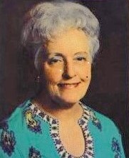
Virginia Livingston, MD (1906-1990)
In this part of the story on cancer I can only ask you to look into the face of this wonderful women and not only see a natural loving kindness but also realise that what she had to endure right up to her death in 1990 was unbelievable red tape and ridicule.
Virginia Livingston never gave up on her cancer research and although she continued to learn so much from fellow peers she would have to be the only possible women that actually understood cancer in its many forms and but also knew how to cure it.
This is an amazing story of the woman who put cancer in permanent remission and in doing so not only cured it but also exposed cures for so many other deadly diseases that all had one thing in common “They all had bacterium as there nucleus, all were possibly infectious and all could be cured”
Don’t you find it sad that when a person has gained so much respect in the medical profession, written so many medical journals that were praised by all who read them and then suddenly attacked and ridiculed because she had found a cure for cancer!
All the respective governments, health departments, oncologists, cancer foundations and the massive pharmaceutical industry suddenly went on the attack when they realized that not only had she found a cure but also had the ability to cut their multi billion dollar scam research into “Not finding a cure.”
Hopefully one day this dear lady will receive a well deserved posthumous award for her services to humankind!!
I will first cover her story and then follow up with many other pioneers in the industry who used Virginia protocol in their own research.
Believe me you will be amazed at all the lies and deceit that continue to this day when there has always been a cure, especially when you can identify the cause (Bacterium) which this lucrative industry has covered up for far too long!!
It is time for the truth to come out and time to bring those responsible for this cover-up to go on trial for crimes against humanity……to put it into its true perspective the link between TB- Cancer – HIV AIDS in the Bacterium sense is unbelievable.

It should also be noted that HIV-AIDS was intentionally introduced into the world’s population via Africa…….just think – more than 25 million people have died including half a million Americans and more than 40 million are currently infected……I could extend this story into another western scam called Ebola and no doubt you will be amazed to find out that there is also a cure for that……and how about the damage being done to African children with highly toxic vaccines….but who’s really interested.

I won’t go into who spread HIV AIDS but it is said a certain US military base had perfected Chemical and Biological Warfare…..not forgetting the help of the British in the production of Anthrax and other deadly diseases and gases which were clearly exported around the world and which gave Saddam the ability to poison his own people in Iraq, the Iranians during the Iraq-Iran War and on Coalition Forces during the Gulf Wars….not to mention the labs and the ingredients that were given to him by the west!!!!!
Now back to the wonderful story of Dr. Virginia Livington as provided by many of her associates and admirers:
Dr, Virginia Livingston with another pioneer Dr. Alan Cantwell
The above photograph was taken in 1981 nine years before Dr. Livingston’s death….she continued her research and her fight with the various authorities right up to her passing and who actually commenced to issue a subpoena the day after her death…….
The following is an extract –
[1983] The Conquest of Cancer: Vaccines and Diet by Virginia Livingston Wheeler M.D. – Edmond G. Addeo
Virginia C. Livingston–Integrating Diet, Nutritional Supplements, and Immunotherapy
The story of Virginia C. Livingston, a physician who died in her late eighties in 1990, is at once dramatic and prototypical of those who venture off the path of mainstream cancer medicine. After undertaking some exacting research, she claimed that she had discovered a microbe that caused cancer and then developed a vaccine that she claimed would help control that microbe. Whether or not her claim has any validity has not yet been fully evaluated, although it has been categorically dismissed by mainstream medicine.
In addition to her vaccine, Livingston also developed a multifaceted nutritional, medical, and immuno supportive program, which can be traced back in part to the German naturopathic tradition of Max Gerson and Josef Issels. She can therefore be considered a “second-generation” nutritional cancer therapist, especially since fully half of her program is nutritional in content. Livingston’s treatment is still being offered at the Livingston Clinic in San Diego.1
Livingston’s Biography
Virginia Livingston started her medical career as one of the pioneering women physicians of her time. Her great-uncle and father were both physicians, and her father was one of the early members of the American College of Physicians. She was one of only four women to receive her M.D. from New York University in 1936 and was appointed the first woman resident physician at a New York hospital–the prison hospital for venereally infected prostitutes.2
While at the prison hospital, she became interested in tuberculosis and leprosy, which were being treated in nearby infectious disease units. As a school physician a few years later, she became interested in scleroderma, a degenerative disease of the skin and tissue that Livingston came to see as related etiologically to tuberculosis, leprosy, and cancer.
Livingston found that a red dye staining technique revealed numerous “acid-fast” organisms (organisms that stained when exposed to the diagnostic dye) in scleroderma. These organisms, she believed, were similar to those found in leprosy and tuberculosis. Because she saw scleroderma as similar to cancer, she began to wonder whether she might not find similar organisms in cancer tissue. “At this point,” she writes, “I reasoned that perhaps scleroderma was a kind of slow cancer. I decided to begin examining cancer tissues with the same method. … Upon examining all kinds of cancerous tissues … I found that a similar microorganism was present in all of them.”3
Livingston then made contact with Eleanor Alexander-Jackson, M.D., of Cornell University, who had found that the tubercle bacillus undergoes many changes in shape, and hence is “pleomorphic” (able to change shape and size.) At that time, cancer was thought to be caused by a virus, and the technique for differentiating a virus from a bacillus was to see whether it passed through a special filter; viruses are much smaller than bacilli and could pass through the filter. Her association with Alexander-Jackson led Livingston to conceive of the acid-fast organisms she saw in scleroderma, leprosy, tuberculosis, and cancer as a family of pleomorphic organisms that sometimes assumed very small forms similar to viruses and at other times had large forms similar to bacilli.4
Against considerable odds, Livingston was able to develop a research program to explore this world-class research hypothesis: a family of microbes–able to change dramatically in size and shape–were responsible for the development of cancer, tuberculosis, leprosy, and scleroderma. At a time when women physicians were scarcely welcome in leading roles in cancer research–much less as champions of major breakthrough concepts–she created the Rutgers-Presbyterian Hospital Laboratory for the Study of Proliferative Diseases associated with the Bureau of Biological Research of Rutgers University.
She received funds from the American Cancer Society and an impressive group of foundations and medical laboratories. “The next few years at Rutgers,” Livingston writes, “were to be the most significant period of my work in cancer research. Our research team was enthusiastic that our work would prove once and for all that the Progenitor cryptocides [or PC, the name she would later give the organism she believed she had discovered] microbe was the cause of cancer and that a vaccine could be made to defend against it.”5
Alexander-Jackson left Cornell to work with Livingston at the new laboratory, and they built a small research team. In 1950, she and Alexander-Jackson published a paper in the American Journal of Medical Sciences, which was co-authored by four others including James Hillier, developer of the electron microscope and head of electron microscopy at RCA Victor Laboratories in Princeton, and John Anderson, head of the department of bacteriology at Rutgers and a noted histologist and pathologist. In this paper, they described how Koch’s postulates (“the accepted foolproof method of proving the cause of a disease”) could be satisfied in the case of P. cryptocides. Pure P. cryptocides cultures were obtained from both human and animal cancers and injected into animals capable of being infected. Disease areas then developed resembling those from which cultures were taken. Pure cultures were then re-isolated from the infected animals. “Koch’s postulates,” Livingston writes, “were fulfilled to the satisfaction of our entire group and to that of our biology superiors at Rutgers.”6
Livingston had thus demonstrated to her own satisfaction, and to that of some colleagues, that she had isolated a microorganism that caused cancer in both animals and humans. Needless to say, for Livingston and anyone who credited her discovery, this was a historic accomplishment. “The next step,” she says,
was to prove that the cancerous growth was not the whole disease. For more than one hundred years people like Rudolf Virchow thought that cancer cells themselves were parasites within the body. He did not understand that the small coccuslike granules he saw dividing in the cancers were not the development of daughter cells within mother cells, but instead they represented the true intracellular parasite that was the causative agent. … The whole truth may be that the parasite within the cancer cell transforms the normal cell into a sick cell that cannot mature by normal cell growth processes. In other words, the tumor is not the disease.7
Her claims did not, however, find approval in the medical community. In 1953, a spokesman for the New York Academy of Medicine, Dr. Iago Gladston, discounted her claims, echoing the attitude of most of the medical community. “This is an old story,” he said, “and it has not stood up under investigation. Microorganisms found in malignant tumors have been found to be secondary invaders and not the primary cause of malignancy.”8
That Livingston’s bold thesis did not find approval in the medical community is not surprising, but the strong opposition to allowing her to continue her efforts to develop and defend her thesis was scientifically unconscionable. As a result, Livingston’s laboratory in New Jersey was forced to close in 1953 due, according to her, to the efforts of leading researchers at Memorial Sloan-Kettering Cancer Center in New York opposed to her research.9 Deeply disappointed, Livingston moved to California to live near her family. But in Europe, and within a small faction of the microbiology research community in the United States, interest in theories similar to hers continued to develop.
Livingston became an Associate Professor of Microbiology at the University of California in San Diego to continue her research. In 1969, she and Alexander-Jackson and their colleagues presented a group of papers at a New York Academy of Sciences meeting on “microorganisms associated with malignancy.” Some of the articles were published in the Annals of the New York Academy of Sciences.10
Livingston viewed P. cryptocides as what she called an “obligate symbiont”: an organism necessarily present in all human cells, in fact one that plays a vital role in all reproductive life, including fertilization and pregnancy and the development of the cancer cell.11 But this obligate symbiont is susceptible to a malignant transformation and proliferation in disease states, especially those that depress normal immune function. She saw cancer as an immunodeficiency condition caused by environmental toxins and inadequate diet.12
In other words, in the classic dispute between researchers who give primary importance to the infectious agent in a disease and researchers who believe that the infectious agent only takes hold in an organism weakened by poor nutrition, toxins, or other stressors, Livingston came down in the middle. The agent did, she believed, play a critical role: it could be isolated and a vaccine effective for the prevention of cancer and modulation of existing cancer could be developed. But the weakened terrain of the organism was also critical, for it provided the depleted environment in which the infectious agent took on pathological shape and multiplied out of control.
In 1990, the Office of Technology Assessment summarized the prevailing attitude about Livingston’s work within the research establishment:
[Dr. Livingston has] little support, outside of a few researchers, for her belief that the different microbes observed in the tissue and blood of cancer patients are actually different forms of the same microbe. At present, no independent evidence exists to corroborate her contention that the microbial forms are related to each other as different forms of a single, pleomorphic organism. Evidence does show that the bacterial culture Livingston isolated is not a new and unique species as claimed: P. cryptocides supplied by Livingston were identified as different species of the genus Staphylococcus and Streptococcus. The issue of isolating bacteria of any kind from tumor tissue and urine of cancer patients, however, is generally not disputed, since many groups of researchers have reported isolating various species and strains of bacteria from such sources. Some of these bacteria have also been showed to undergo morphogenic alterations characteristic of cell wall deficient (or pleomorphic) bacteria.13
It should be noted that the same author has also written another book about Dr.Livingston:
The Woman Who Cured Cancer: The Story of Cancer Pioneer Virginia Livingston-Wheeler, M.D. and the Discovery of the Cancer-Causing Microbe
by Edmond G. Addeo

In Part 4 I will continue to reveal the incredible work carried out by Dr. Virginia Livingston and work carried out by other medical pioneers who supported not only Dr. Livingston Protocol but also agreed with her research into the connectivity between TB – Cancer-HIV AIDS and many other bacterium based diseases.

Where does all the money go – “God Know’s
It will become clear to you all that governments – health departments – oncologists – cancer foundations/charities and the big pharmaceutical industry have indeed created their own scam in raising billions of dollars in “Not Finding A Cure For Cancer.”
Peter Eyre – 0840 Local Time – 15/6/2017
Middle East Consultant – Political Analysis – Investigative Journalist – Broadcaster
Is There A Cure For Cancer ? You Bet There Is! – Part 2

Lets start by opening up a can of worms and expose the worldwide cancer scam

One of the biggest investment banks in the world
In my last article I covered the basic understanding of what cancer actually is and how it is related to many other serious diseases all of which have Bacterium as their nucleus.
We must fully understand that our respective governments, health departments, oncologists, cancer research institutes, cancer charities, the very lucrative pharmaceutical industry and their “Zionist Controlled Media” are literally making a killing (both financially and physically speaking) in their multi billion dollar scam “In not wanting to find a cure for cancer.”

Who controls the pharmaceutical industry & World Health (WHO) can kill us all
What we know so far and the factual connection between many deadly diseases is that they not only have something in common but they can all be cured by the application of simple medical logic!
Imagine being a cancer victim having suffered their extremely painful and expensive chemotherapy and being pumped up with untold concoctions of medication and morphine only to be told there is nothing more we can do for you!
Imagine being lied too from day one that your cancer is not what they think it is and that it is a bacterium similar to Tuberculosis and although contagious can be cured!
Imagine having multiple operations on your leg, pins or plates inserted and then after many years being told it has become badly infected and that gangrene has set in and now consequently you will have to have your leg amputated when once again a simple medical procedure could have saved it!
Practically speaking it all comes down to simply accepting that bacterium is the root cause of many of today’s more serious diseases and thus if one treats it as a bacterium, identifies that bacterium and reintroduces that bacterium back into the patient you are basically creating an anti venom in much the same way as a snake bite!
You would be shocked if you actually knew the bacterial connection between many of what the scumbags call deadly diseases and how so many innocent victims have passed away (in their millions) when they could have been saved by treating the root cause as bacterium based!
Let’s now link some of these so called deadly diseases, connect the dots and prove that we are all being truly conned in their analysis whilst at the same time being ripped off for medication that we did not require had their analysis been correct in the first place!

We are being ripped off an arm and a leg
The following is an initial list of terrible diseases that have a common bacterium link:
Leprosy, tuberculosis, cancer, scleroderma, Raynaud’s syndrome with a strong possible connection to AIDS.
Couple the above to another relatively unknown treatment called “Phage” for other deadly bugs such as MRSA or badly infected limbs and tissue etc and one can clearly see that our government, health professionals, consultants, oncologist, doctors and pharmaceutical industry either simply do not understand what is causing the problem or alternatively simply refuse to accept for fear of impacting on their multi billion dollar industry, their own ex gratia payment or salary top up etc.
To give you all some idea of the alleged success rate lets look at some figures in regard to cancer treatment that was carried out some time ago by a certain doctor who had been working on cancer for decades and who’s initial work was applauded in her many medical journals only to be ridiculed and closed down when they realized she may well have a cure for cancer…….such is the state of affairs in this very lucrative practice in pretending to find a cure whilst raking in billions of dollars but never finding one!
62 random patients were chosen for her trial whilst accepting that cancer was bacterium based and could be treated with bacterium extracted from each patient, cured in the lab and re introduced back into the patient. This extraction was from the tumor, tissue, gland, blood or urine…….remembering another false declaration by the industry that “Blood is not sterile.”
The 62 patients consisted of the following (Included were 17 patients officially diagnosed as terminal):
The 62 cases included 21 breast cancer patients of various types, 5 lung cancer patients, 3 uterine cancer, 3 ovarian cancer, 6 colon cancer, 6 melanoma, 2 basal cell skin cancer, 3 prostate cancer, 2 kidney cancer, 1 pancreatic cancer, 1 pelvic cancer, 1 esophageal cancer, 1 larynx cancer, and 6 Hodgkin’s disease patients.
In summarizing these cases, she officially claimed, “an examination of the sixty-two random cases shows that our success rate has been 82%. Considering the patients we called inconclusive but for whom we were able to be of some help, it is over 90%”
I also understand that she also drew attention to the fact that her success rate would have been higher had some of them not been treated by chemotherapy or radiation which directly attacks the immune system that is so vital in the recovery of patients!

One can see that our governments, departments of health and the thousands of specialists employed in the above and the pharmaceutical industry really do need to be re educated and exposed for their Weapons of Mass Deceit (WMD) and in some cases placed before the court system to be tried for what they are “Deceitful Imbeciles.”
If you think that this is bad wait till I uncover the same deceit in regard to the world’s vaccination programmes promoted by the New World Order and its puppet the United Nations/WHO under UN Agenda 21 and its replacement programme UN 2030 Agenda to depopulate the world by as much as between 80-90% of current population “by whatever means” and how the ingredients and adjuvants in those vaccines are causing death, maiming, mental deterioration, dumbing up and infertility to our babies, very young and our teenagers…….all of which has been approved and countersigned by our respective governments……some of whom are directly or indirectly benefiting financially by their own investments!!!!


Part 3 will be a more technical brief to reveal the fine work carried out by many superb doctors, who they were, what they found and how they were treated by the above mongrels and finally show that “a cure for cancer and other deadly diseases does exist.”
Peter Eyre 0930 local time 13/6/2017
Middle East Consultant – Political Analysis – Investigative Journalist – Broadcaster
Is There A Cure For Cancer ? You Bet There Is! – Part 1

Over many many years I have tried to understand why they have not found a cure for cancer when billions of dollars continue to be pumped into the many research institutes each and every day throughout the world without a ray of hope being given for the millions of innocent victims who trust their governments, hospitals, cancer clinics, oncologists and cancer organisations in saving them from this terrible disease .
Historically many gifted professors and doctors have hit the cause of cancer on the head but in doing so have been immediately silenced by governments health departments. pharmaceutical companies or cancer institutes by either ridicule or simply having their licence revoked or their potential cure shelved by those who make far more money in not finding a cure!
Why would anyone in their right mind wish to cause so much pain and suffering to the victims of cancer when many extremely talented doctors had looked upon cancer in a totally different light and provided potential treatment that did not harm the body, as radiation and chemotherapy do.

Who in their right mind would allow the bodies natural immune system to be bombarded with radiation and chemotherapy whilst at the same time giving their patients high doses of morphine to a point that one would have to ask the question as to what actually killed the patient in their final days.


How would you feel if you had cancer or your loved one had cancer and were told that the existing understanding of the origins of cancer were all wrong and that all those mentioned above were covering up the real truth as to what is cancer or what causes cancer to develop?
How would you feel if you found out that cancer was closely linked to other serious illness’s such as Tuberculosis (TB) or HIV AIDS etc and was bacterium based and that a potential cure was available?
How would you feel if you were told that HIV AIDS was first intentionally introduced into the world’s civilian populations by the US Military and had no connection to monkey’s or apes?

I know it did not come from me so how come you have become so gullible you believe such bullshit?
So lets now look at cancer from a different perspective and think of it as being bacterium based and accordingly consider that it can now be passed on to someone else i.e. infectious but also fully understand that it is also curable!
Some very notable researchers, who have had their achievements printed in medical journals, came across some very unusual findings in their respective laboratories, suffice to say they all agreed that cancer was indeed a bacterium!
It was during my years of research that I also came across the use of “Phage” to combat some very serious conditions such as MRSA that now exists in many of the world’s hospitals and for which there was no apparent cure……….but there is!
Let me put this to you in a very simple way:
If you are bitten by a snake the hospital identifies the culprit, finds the same venom and use it as a anti venom to put back into your already contaminated body to fight the same venom or bacteria.
In a nutshell if we are infected with bacterium in any form then if one is able to find that same bacteria either by extraction from the victim or find the same bacterium in its natural environment, take it back into the lab and create a microbiological culture and eventually re introduce that bacterium back into the patients body makes perfectly good sense!
One must also keep in mind the United Nations Agenda 21 and its replacement UN 2030 Agenda regarding mass depopulation by whatever means (which has been countersigned by many countries) is clearly targeting the world’s population via their approved fast track vaccination programmes, unwillingness to cure treatable diseases and using measures that are clearly detrimental to ones health!
In Part 2 we will further explain just how closely linked many of today’s diseases are and prove that their nucleus is bacterium based along with the research carried out by some notable medical practitioners who’s experience cannot be challenged!
Peter Eyre
Middle East Consultant – Political Analysis – Investigative Journalist – Broadcaster
